
Take the next step
Carry the full power of our ecosystem in your pocket. Transform your habits and reach your goals today, right from your iPhone.

Or scan the code to install:

Carry the full power of our ecosystem in your pocket. Transform your habits and reach your goals today, right from your iPhone.
Or scan the code to install:


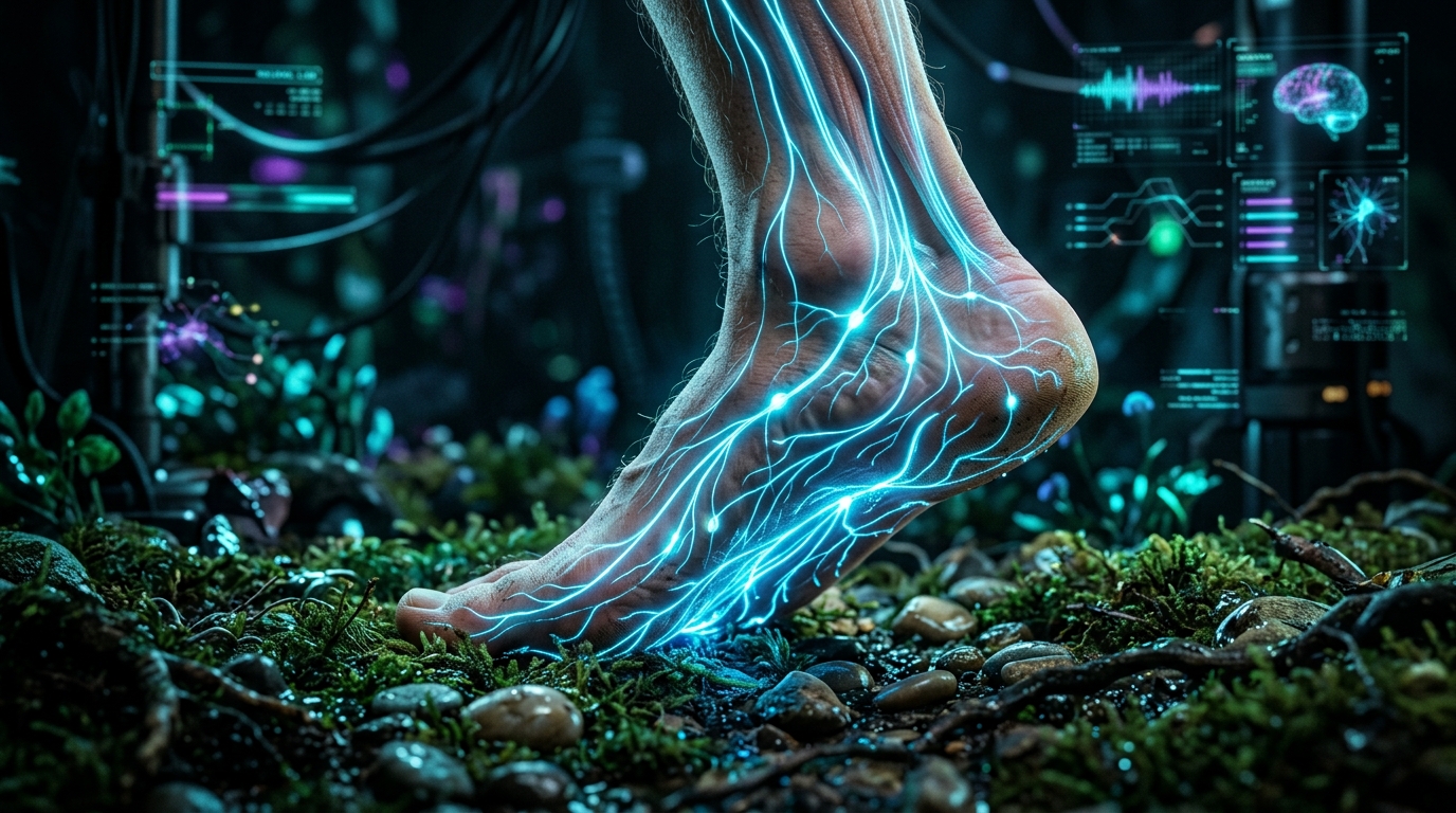
Pie
Obligate bipedalism is one of the most defining and biomechanically complex evolutionary milestones of the human species. Throughout millions of years of adaptive evolution, beginning with early hominids and perfected in the genus Homo, human anatomy was sculpted by environmental demands to withstand massive dynamic loads, provide shock absorption, store elastic energy, and act as a rigid lever system indispensable for efficient propulsion. The endurance running hypothesis suggests that humans evolved not to be the fastest mammals over short distances, but to become persistence hunters, capable of tracking and exhausting prey through long-distance running thanks to a superior thermoregulation system and a foot architecture extraordinarily efficient at conserving energy. In this evolutionary context, the human foot developed by interacting directly with terrain irregularities, without any external assistance, building an intrinsically self-sufficient musculoskeletal and neurological system.
However, in recent history, and particularly following the industrial revolution and the rise of athletic footwear in the 20th century, the development of modern footwear has introduced artificial biomechanical alterations that deeply challenge and subvert this meticulous evolutionary design. The predominant paradigm in the footwear industry has maintained that the human foot is an inherently fragile or flawed structure requiring constant orthopedic support, extreme cushioning to mitigate impact, and an elevated heel to facilitate gait transition.
The contemporary debate in movement science, sports medicine, podiatry, and clinical biomechanics is increasingly centered on the evident dichotomy between traditional footwear—characterized by elevated heels or “drop,” thick layers of viscoelastic cushioning, rigid arch supports, and aesthetically narrow toe boxes—and minimalist footwear, whose design seeks to interfere as little as possible with the natural movement and kinematics of the foot. Current scientific research and three-dimensional kinematic analyses overwhelmingly demonstrate that highly technological footwear not only alters natural walking and running patterns but also generates profound morphological, structural, and neuromuscular changes.
The chronic use of traditional footwear essentially acts as a prolonged orthopedic cast worn daily. Like any immobilized extremity, this mechanical isolation immobilizes the joints of the foot, severely weakens the intrinsic musculature through disuse, and pathologically alters the transmission of forces along the entire kinetic chain, projecting mechanical dysfunctions from the ankle joint up to the knees, hips, and lumbar spine. Conversely, the scientific philosophy behind minimalist or “barefoot” footwear is based on rigorous biomimicry: respecting natural movement patterns and allowing the foot’s intrinsic anatomical structures to actively perform the work of shock absorption, stabilization, and propulsion for which they were biologically designed and optimized.
This comprehensive research report deeply, anatomically, and thoroughly analyzes how the biomechanical characteristics of traditional footwear induce severe physiological side effects. It explores how these artificial interventions alter shock absorption, pathologically shorten the Achilles tendon, atrophy musculature, and weaken the human body’s base of support. Even more significantly, this analysis details how this footwear-generated loss of lower limb strength directly correlates with an increased risk of falls, a severe loss of motor independence, and a general, statistically significant reduction in human longevity. Simultaneously, it establishes the biomechanical and neurophysiological foundations that support the benefits of minimalist footwear as a fundamental prophylactic and therapeutic tool for restoring optimal anatomical function, proprioception, and structural resilience.
To fully understand the detrimental effects and mechanical limitations imposed by restrictive footwear, it is first absolutely imperative to dissect and analyze the sophisticated three-dimensional architecture of the human foot. Far from being an inert anatomical block or a simple passive base upon which the skeleton rests, the foot is a dynamic, adaptable, and highly reactive organ composed of an intricate bony, articular, and cartilaginous network.
Bone, Joint, and Morphological Organization The human foot is composed of 26 bones, meaning that both feet house approximately one-quarter of all the bones in the entire human body. These bones are connected by 33 distinct joints and stabilized by a complex network of over 100 ligaments, tendons, and muscles. Structurally and functionally, the foot is divided into three main segments that act synergistically during the gait cycle: the hindfoot, midfoot, and forefoot.
The hindfoot consists of the largest bone in the foot, the calcaneus (heel bone), and the talus, which forms the ankle joint by connecting the foot to the tibia and fibula of the lower leg. The evolutionary morphology of the human talus shows flexible profiles adapted for walking long distances over uneven terrain, a characteristic restricted by the constrictive footwear of post-industrial societies.
The midfoot is the segment responsible for forming the plantar arches. It is composed of five smaller bones: the navicular, the cuboid, and the three cuneiform bones (medial, intermediate, and lateral). These bones fit together like the stones of a Roman architectural arch, providing structural stability without relying on central pillars.
Finally, the forefoot includes the five metatarsal bones and the 14 phalanges that make up the toes. Each toe, except for the hallux (big toe), consists of three phalanges (proximal, middle, and distal), while the hallux, crucial for propulsion and balance, has only a proximal and a distal phalanx. The metatarsophalangeal joints in the forefoot are the exact inflection point over which the body pivots forward with each step.
The Musculature of the Foot: The Active Subsystem and the “Foot Core” The structural and functional integrity of the foot is orchestrated by its musculature, which is anatomically divided into two categories: extrinsic muscles and intrinsic muscles. Extrinsic muscles originate in the compartments of the lower leg (calf and shin) and insert into the foot via long, robust tendons, being primarily responsible for the large movements of ankle dorsiflexion, plantar flexion, inversion, and eversion.
However, the intrinsic foot muscles (IFM) are the true protagonists of local stability and arch support. These muscles have both their origin and insertion strictly confined within the structure of the foot itself, meticulously organized into multiple plantar layers. Key muscles in this group include the abductor hallucis (AbH), which originates at the calcaneal tuberosity and abducts and flexes the first toe; the flexor digitorum brevis (FDB), responsible for flexing the second through fifth toes; the abductor digiti minimi (ADM); and the plantar and dorsal interossei muscles.
Analogous to how deep abdominal and lumbar muscles stabilize the spine (the body’s “core”), the intrinsic muscles of the foot function as the local stabilizers of the “foot core system.” Their primary function is not to generate large ranges of motion, but to precisely control the deformation of the medial longitudinal arch during dynamic weight-bearing, modulate midfoot joint stiffness, and ensure that plantar pressures are optimally distributed across the metatarsal heads.
The fundamental error of the traditional footwear industry has been assuming that the impact of human locomotion against the ground must be absorbed by external synthetic materials, such as thick foams or air chambers. The biomechanical reality is that the human foot already possesses nature’s most sophisticated cushioning systems, designed specifically to dissipate ground reaction forces and recycle that energy. The two primary mechanical systems that achieve this are the arch-spring mechanism and the windlass mechanism.
The plantar fascia plays an indispensable role in both mechanisms. This is a thick aponeurosis, a band of dense, inelastic connective tissue that extends from the medial tuberosity of the calcaneus in the heel, fanning out across the sole of the foot to insert at the base of the proximal phalanges of all five toes.
The Arch-Spring Mechanism In 1943, researcher Lapidus first modeled the medial longitudinal arch of the foot as a truss model. In this model, the bones of the foot form the upper compression arch, while the plantar fascia and plantar ligaments act as the lower horizontal tension tie-rod connecting the ends of the arch.
During the first half of the gait cycle (initial contact and loading response), when full body weight lands on the foot, gravitational forces attempt to flatten or collapse the medial longitudinal arch. As the arch deforms downward, the plantar fascia and elastic plantar ligaments passively stretch, while the intrinsic foot muscles contract eccentrically (lengthen under tension) to control the speed and magnitude of this descent. This controlled anatomical deformation is the human body’s primary shock absorption mechanism, dissipating kinetic energy that would otherwise travel as a destructive shockwave toward the tibia and knee. By elongating, these elastic structures store an immense amount of strain energy, operating literally as a biological spring.
The Windlass Mechanism If the foot remained solely as a soft, flexible shock absorber, it would be mechanically disastrous for the propulsion phase of gait, as attempting to push the body forward using a soft structure would result in a massive loss of muscular effort. This is where the brilliance of the windlass mechanism comes into play.
Biomechanical analysis shows that during late stance and toe-off, the toes—and most critically, the robust big toe or hallux—dorsiflex (bend upward) as the heel lifts from the ground. As the big toe dorsiflexes, the plantar fascia, anchored to the phalanges, physically winds around the metatarsophalangeal joint, which acts as the drum or axis of a windlass.
This winding action subjects the plantar fascia to tremendous tension, pulling the calcaneus forward toward the metatarsal heads. This physically shortens the base of the foot, dramatically elevates the longitudinal arch, and firmly locks the midfoot bones (Chopart’s joint). As a direct result, the foot transforms in milliseconds from a soft, mobile adaptor (perfect for cushioning) into an incredibly rigid, supinated propulsion lever, allowing for optimal propulsive energy transfer that eliminates any reliance on artificial footwear cushioning.
However, the uninterrupted use of rigid traditional footwear from childhood prevents the toe joints from achieving the necessary dorsiflexion, stopping the windlass mechanism from developing properly and operating normally, which permanently compromises the one-to-one coupling between toe flexion and arch tensioning.
The vast majority of traditional footwear—ranging from formal dress shoes and work boots to modern, highly cushioned running shoes—shares three anatomical characteristics that severely alter natural physiology: heel elevation, a narrow toe box, and a thick layer of cushioning. Far from being benign, these design features introduce chronic pathomechanics.
The Impact of Heel Elevation (Drop): Achilles Tendon Shortening and Loss of Dorsiflexion The “drop” of a shoe is the net height difference between the heel and the toe. In conventional athletic footwear, this drop typically ranges from 6 to 12 millimeters, while in formal footwear or high heels, the elevation is significantly greater. Keeping the body in a posture where the heel is perpetually elevated above the forefoot places the ankle joint in a constant state of forced plantar flexion.
The physiological effects of this chronic plantar flexion are profound and detrimental. In the short term, this position is bioenergetically inefficient, causing excessive overlap of actin and myosin filaments in the calf muscle fibers (gastrocnemius and soleus), forcing them to operate outside their optimal length-tension range. To compensate for this mechanical inefficiency, the neuromuscular system undergoes a long-term maladaptive plastic adaptation: the gastrocnemius muscle fascicles physically shorten to reposition the sarcomere overlap back to its new, shortened operating regime.
Simultaneously, the Achilles tendon, the strongest fibrous band in the body connecting the calf to the heel bone, adapts to this lack of elongating tension by contracting and becoming abnormally stiff and thickened. Ultrasound and kinematic research have precisely measured this deterioration; it has been shown that for every 1,000 daily steps taken in shoes with an elevated heel, the structural stiffness of the Achilles tendon experiences an alarming increase of 9.2%.
This chronic adaptation drastically reduces the ankle joint’s active range of motion (ROM), specifically severely limiting dorsiflexion, which is the ability to bring the foot toward the shin. Limited ankle dorsiflexion immediately alters the kinematic movement pattern of the entire lower extremity during gait, decreasing the body’s ability to naturally propel itself forward. When an individual with this shortening attempts to walk barefoot or wears flat shoes, they immediately experience discomfort, tension, and pain, as their tissues have lost the capacity to stretch to their evolutionary anatomical length. Furthermore, this lack of mobility exposes the tendon to abnormal stresses during athletic activities, being the primary underlying cause of Achilles tendinitis and tendinosis, conditions characterized by acute inflammation and microscopic degeneration of the tendon’s collagen fibers.
The Narrow Toe Box: Destruction of the Plantar Tripod Stability The natural foot in its evolutionary state is significantly wider at the tips of the toes than at the metatarsal region. However, fashion aesthetics have dictated that modern footwear converge into a funnel shape, crushing and confining the toes into a narrow, tapered toe box.
This chronic physical constriction nullifies one of the body’s most vital stability mechanics: toe splay. When a healthy foot makes contact with the ground and bears weight, the toes naturally expand outward to significantly widen the base of support, stabilize lateral balance, and evenly dissipate impact forces across the metatarsal heads.
Prolonged confinement of the toes within a rigid shoe severely weakens the intrinsic foot muscles, particularly the abductor hallucis muscle. When the big toe is forced into lateral deviation (toward the other toes), the first metatarsal deviates in the opposite direction. Epidemiological research indicates that wearing narrow footwear is the primary and fully modifiable etiological factor in the development of Hallux Valgus (bunions), a painful structural joint deformity affecting a staggering 19% of the global population. A foot with imprisoned toes loses its capacity to serve as the fundamental stabilizing tripod, which dangerously elevates plantar pressures in inappropriate regions of the forefoot (causing metatarsalgia) and compromises the individual’s directional agility.
The Paradox of Artificial Cushioning and Muscle Atrophy The selling point of traditional athletic footwear is that the thick viscoelastic midsole attenuates repetitive stress by passively absorbing impact forces. However, scientific studies in advanced biomechanics reveal that this premise is flawed due to what is known as the “cushioning paradox.”
The presence of a thick layer of foam dramatically interferes with the sensory feedback the foot sends to the brain. By not accurately perceiving the hardness of the ground, the nervous system suppresses its natural biomechanical protective instincts. Users of cushioned footwear pathologically lengthen their stride (overstriding), land with a straight knee ahead of their center of gravity, and generate a violent initial contact with the heel (rearfoot or heel strike), mistakenly trusting that the foam will dissipate the energy.
Research published in Scientific Reports evaluated this paradox through three-dimensional analysis of runners using highly cushioned shoes versus thin-soled footwear. The results were counterintuitive but biomechanically irrefutable: when striking the pavement in thick shoes, individuals flexed their knees significantly less. Because the thick foam platform collapsed and created instability under the foot, the brain induced a compensatory response in which it made the entire leg mechanically stiffer to maintain balance. This excessive lower limb stiffness during landing caused transient impact loading rates to travel at greater speeds and magnitudes through the bone tissue compared to wearing a thin-soled shoe.
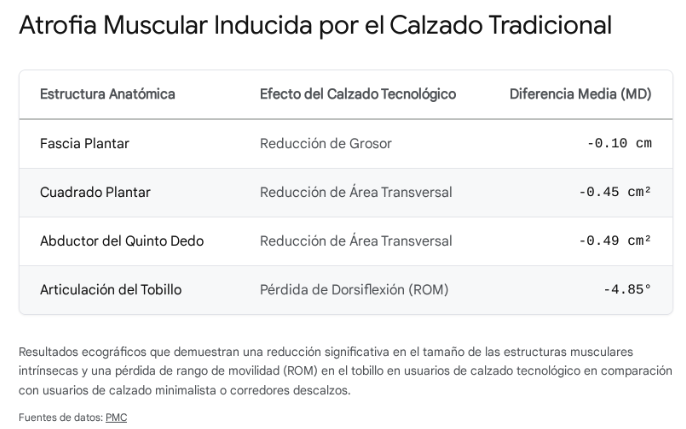
Beyond the increase in shock forces, constant cushioning and the rigid arch support built into the midsole wreak havoc on the foot’s muscular health. If an anatomical structure is not used because an external agent (the shoe’s orthopedic support) performs its function, it degenerates. Traditional technological footwear inactivates the intrinsic foot muscles, resulting in quantifiable muscle atrophy.
atrofia muscular por el calzado
Evidence obtained through ultrasound measurements demonstrates that runners who exclusively use technological footwear exhibit statistically significant reductions in plantar fascia thickness and in the cross-sectional area of multiple intrinsic muscles compared to populations that run barefoot or habitually wear minimalist footwear.
The human body does not operate as a collection of independent parts, but under the biomechanical guiding principle of an integrated and mutually dependent kinetic chain. In this model, an anatomical or mechanical dysfunction at the base of support—the foot—never remains confined there, but provokes massive compensatory perturbations that relentlessly ascend through the musculoskeletal system.
When traditional footwear “casts” and weakens the foot, completely disabling the natural cushioning function of the muscular plantar vault, the arch-spring mechanism, and the windlass mechanism, the immutable laws of physics come into play. The kinetic energy and powerful impact forces generated by walking and running do not magically disappear due to the lack of a functional foot; instead, all this unabsorbed vibratory and shear energy is violently transferred up the chain, mercilessly punishing the higher structural joints: the knee, the hip, and the lower back.
Morphologically, profound weakness of the intrinsic foot musculature prevents the medial longitudinal arch from maintaining its architecture under body weight, resulting in its inward collapse into a posture of severe overpronation. From the kinetic chain perspective, excessive foot pronation obligatorily induces a massive and simultaneous internal rotation of the tibia and femur. This spiraling rotational misalignment asymmetrically increases shear stress on the knee’s collateral ligaments, grinds the meniscal cartilage, and misaligns patellar tracking within the trochlear groove.
Unlike the complex ball-and-socket joint of the hip or the multidirectional ankle joint, the knee is primarily a simple hinge joint, biologically designed to flex and extend in a sagittal plane, not to resist extreme torsional forces. Forcing the knee joint to act as the primary absorber of blunt force and rotational impact that should have been attenuated and dispersed by the foot’s sophisticated anatomy is the main precursor to degenerative knee osteoarthritis and patellofemoral pain syndromes.
Simultaneously, global body posture undergoes a dramatic transformation due to the geometric profile of traditional footwear. The artificial heel elevation caused by the drop mechanically tilts the body’s entire axis forward, significantly altering the individual’s center of gravity. Since maintaining a full forward tilt would cause a person to fall flat on their face, the central nervous system commands immediate biomechanical compensations: the individual must create an anterior pelvic tilt (deep pelvic anteversion) that rotates the hips backward.
To keep the torso upright and the line of sight parallel to the horizon with this tilted pelvis, an extreme compensatory increase in the lordotic curve of the lumbar spine is forced. This chronically sustained spino-pelvic misalignment pathologically shortens the robust erector spinae muscles and drastically increases asymmetrical compression forces on the posterior portion of the intervertebral discs and the small lumbar facet joints. This is how the simple act of elevating the heel in footwear lays the insidious mechanical groundwork for the chronic development of debilitating lower back pain, radiculopathies, and disc herniations over years of uninterrupted use.
Understanding the human foot exclusively as a mechanical collection of pulleys, springs, and levers is to settle for a reductionist analysis. Fundamentally, the sole of the human foot is a powerful neurosensory organ and the brain’s primary tactile extension for interaction with the physical world. The glabrous (hairless) skin of the sole is densely and intensely innervated by a complex network of specialized cutaneous mechanoreceptors.
Neural mapping studies have identified over 104 extensively documented mechanoreceptive afferent units on the plantar surface. These sophisticated tactile sensors are categorized into two functional groups. On one hand, there are fast-adapting receptors (FA I and FA II, including Pacinian and Meissner’s corpuscles), which make up approximately 71% of the total and are extremely sensitive to high-frequency microscopic vibrations, fast stroking, and sudden transitions in impact forces. On the other hand, slow-adapting receptors (SA I and SA II, corresponding to Merkel discs and Ruffini corpuscles), representing the remainder, are calibrated to relentlessly monitor constant static pressure, skin stretch thresholds, and the topographical distribution of body weight.
The synergistic integration of information from this mechanoreceptor network allows the entire plantar surface to function neurologically as a continuous “dynamometric map” for the intricate control of human balance. Proprioception, which is the deep, internal kinesthetic sense of the body and limbs’ position and movement in space, intrinsically and vitally depends on this vast inferior neural network.
This valuable tactile and afferent sensory information travels at millisecond speeds to the spinal cord and central nervous system, allowing the intrinsic foot muscles, in conjunction with the large global stabilizers of the legs and trunk, to make automatic muscular micro-adjustments via motor reflex arcs to counteract gravity and maintain flawless upright posture over dynamic, uneven surfaces. During active locomotion, this neural sensitivity alerts the brain to terrain hardness, incline, and texture, enabling it to instantly modify limb stiffness, adjust the articular angle of attack, and deploy protective reflex responses.
The use of thick layers of synthetic EVA soles, air chambers, and gel insoles in traditional footwear artificially masks and absorbs almost all of this fine sensory feedback, literally deafening the crucial neurological dialogue between the ground’s topography and the cerebral cortex. By chronically depriving the somatosensory nervous system of these high-definition tactile stimuli, the body loses its capacity to preemptively and subtly modulate limb dynamics in anticipation of impacts and irregularities. In response to this footwear-imposed sensory deprivation, people develop clumsy motor patterns, pronounced alterations in center-of-gravity control, and mechanically much less coordinated gait.
Minimalist footwear design immediately solves this neurological deficit. By employing a millimeter-thin sole devoid of spongy cushioning, the tactile barrier is eliminated, and the massive flow of the foot’s natural exteroceptive and proprioceptive information to the brain is restored. This sensory reconnection deeply enriches the quality of information processed by the cortical motor areas, stimulating neuroplasticity and refining neuromuscular pathways to optimal efficiency levels. During developmental stages like childhood, this enrichment of somatosensory input is fundamental; unmitigated plantar contact allows for superior body mapping in the brain, fostering the intrinsic development of advanced motor agility, cross-coordination, and the instinctive automation of fall-prevention postures. In adults and elderly populations, reintroducing this plantar vibratory sensitivity allows intricate muscular synergies to recalibrate and operate as biology dictated, instantly improving subconscious postural alignment, hastening stabilizing reflex responses, and facilitating firm traction with the environment that effectively protects against accidental trips and sprains.
In glaring contrast to the overwhelming number of corrective components, stabilizing wedges, and synthetic chambers of the modern footwear industry, minimalist or “barefoot” footwear represents an intentional deconstruction and a calculated return to the design of biological biomechanics. Unlike a redesigned conventional shoe, a true minimalist shoe is strictly defined not by the technologies added to it, but by the barriers it removes, and it must meet four uncompromising functional and structural characteristics to genuinely simulate barefoot dynamics while providing a minimum degree of protection against extreme temperatures or sharp external elements:
Zero-Drop: There is total and mathematical leveling between the heel plane and the forefoot plane (0 millimeters height difference), allowing gravity to act on body biomechanics in a physiologically identical manner to standing on a natural flat surface, preventing lordotic alterations and Achilles shortening.
Millimeter-Thin and Highly Flexible Sole: Construction of a plantar base as thin as materially possible (frequently between 3 mm and 5 mm) that not only maximizes terrain proprioceptive sensitivity but also, being mechanically flexible in all directions (capable of being rolled up on itself), allows the foot to articulate uninterrupted through its full range of flexion, torsion, and extension during the entire gait cycle.
Anatomical Last with Wide Toe Box: A perimeter design that faithfully replicates the asymmetric silhouette of the human foot, with the toe segment of the shoe being substantially wider than the heel, eliminating any lateral compressive force vectors and ensuring full three-dimensional expansion (splay) of the phalanges during directional weight transfer.
Absolute Absence of Structural Cushioning and Arch Support: Elimination of robust medial fascia stabilizing wedges and thick viscoelastic (EVA) midsoles, imperatively returning to the intrinsic bony, articular, and ligamentous network of the human foot’s longitudinal arch its non-transferable function as the sole dynamic spring system and biological impact attenuator.
The clinical transition and sustained adoption of minimalist footwear, when done measuredly and progressively to prevent tissue overload, trigger profoundly positive muscular, morphological, and biomechanical structural adaptations in the body. Controlled studies invariably demonstrate this phenomenon. In a rigorous four-week randomized clinical trial, researchers prescribed a gradual, monitored walking program using minimalist footwear in adult subjects, with methodical volume increments from 3,000 to 5,000 steps per day.
Findings revealed highly significant anatomical and postural improvements. Primarily, minimalist interventions actively reversed the degenerative atrophy processes generated by traditional footwear, resulting in a notable increase in cross-sectional area and maximum contractile strength of vital intrinsic foot muscles, including the abductor hallucis (AbH) and abductor digiti minimi (ADM). This potent redevelopment and strengthening of foot core muscle hypertrophy demonstrated a quantifiable impact on static architecture: it mechanically stabilized overall foot posture, statistically reducing pathological degrees of excessive overpronation and actively repositioning the plantar arch vault closer to a healthy, physiological neutral position, with sustained benefits detectable long after the protocol ended.
From a kinematic and dynamic standpoint, removing the elevated heel frees the gastroc-soleus complex from its chronic restrictions, inducing functional fiber lengthening and allowing the recovery of a wide and powerful arc of ankle dorsiflexion. Likewise, when walking or running in truly minimalist footwear, the user makes an instinctive change in their gait pattern guided by the influx of neurological sensitivity, abandoning the violent initial heel strike with extended knees to adopt a biologically natural stride: shorter, more frequent steps, controlled protective landings on the midfoot or forefoot area, strategically flexing the knees to mitigate tension. This intelligent redistribution of joint moments and angles is not a trivial alteration; it massively reduces the anomalous peaks of transient collision forces and initial braking impact, efficiently unloading the destructive stress that the modern stride deposited unattenuated onto the femorotibial menisci, hip, and lower back, dissipating the load across muscles, fasciae, and tendons conditioned for such work.
The progressive and insidious weakening of the foot’s musculoskeletal structures passively generated by the universal standardization of modern restrictive footwear does not constitute, from any viewpoint, a secondary concern of orthopedic aesthetics or isolated athletic performance; on the contrary, it represents a primary, underestimated epidemiological risk factor that casts a deep, measurable shadow over overall metabolic health viability, functional senescence, resilience to physical traumatic events, and the net longevity of the individual throughout the lifespan.
The architecture of the human skeleton inevitably operates under dynamic vector forces where the performance of the upper strata inexorably depends on the non-negotiable integrity of the base and lower supporting foundations. Therefore, there is an irrefutable, highly statistically reliable biological, metabolic, and clinical correlation between the basal structural strength of the legs, positional stability, the contractile and sensory function of the tiny, thick foot muscles, and the deviation of general mortality rate curves across all etiologies and determining causes.
Quantitative Muscle Strength of the Lower Extremities as a Central Predictive Metric of Global Mortality Risk The overwhelming evidence from recent physiological clinical research has conclusively established across numerous global research consortiums that the quantifiable metric strength of the deep leg musculature serves as an infallible primary macroscopic biomarker and predictor of metabolic vascular health, physical resilience against morbid episodes, and ultimately, the effective length of a human’s chronological life expectancy.
The analysis of multiple immensely large, systematic long-term follow-up cohorts in epidemiological studies revealed that adult individuals situated in brackets with intrinsically higher levels of vigor and muscular robustness demonstrated in the lower pelvic extremities (parameters scientifically evaluated through dynamometry and instruments specifically measuring, among other markers, the maximum isometric power generated in functional tests of knee extension and pelvic hip flexion) consistently experienced, without notable environmental variations, impressively steep and massive reductions in their individual and corresponding population profiles regarding the statistical and factual underlying risk of total recorded mortality.
The profiles outline lethal consequences for weakness. In specific research, mature and elderly adults statistically grouped within the lowest or most deficient functional quartile concerning femoral quadriceps contractile force generation capacity were found to possess an exponentially relative risk of experiencing significantly premature death from natural and external causes compared to their more robust peers, increasing their grim probabilities by a staggering verified interval ranging from an extra 51% to even a severe 65% probability—an undeniable, firm risk meticulously verified after being subjected to rigorous analytical adjustment eliminating any bias and confounding variables such as age, ethnicity, inherited race, underlying chronic inflammatory morbidities, basal anatomical mass index, or history and routines of general physical or sedentary life.
To an even more alarming magnitude in terms of medical risk implications, another meticulous observational cohort study in the Asian nation that analyzed the population at scale formally detected and recorded in its scientific databases corresponding to vital global lethality factors that, unlike simple subtle decreases, those individuals constituting the extreme categorizations corresponding to the groups diagnosed with the lowest level of general muscular anatomical power index of the lower skeletal limbs presented, in the face of any severe diagnosis, an overwhelming parallel rate of skyrocketing general mortality occurrence that was up to a drastic 260% higher in magnitude of proportional percentage damage. This multiplied instances by 2.6 times compared to reported clinical death cases in stark standardized statistical biological comparison with the extended life indices enjoyed by those individuals located and belonging to the vigorous stratum and group ostensibly cataloged as the biomechanically strongest specimens. In monitored patients already presenting prior chronic cardiovascular conditions who had been officially diagnosed suffering from severe chronic painful peripheral arterial pathologies or parallel systemic vascular disease in legs due to atheromatous occlusion (PAD), the specific verified finding regarding weakness in precise indices referring exclusively to the rapid, sustained, gradual decrease detected in the fundamental general metric evaluating the real, biomechanically verified performance of force and power exerted analytically across the pure biological anatomical aspect to achieve and consolidate final physiological articular rear muscle flexion force of the hamstring/knee flexion zone demonstrated fatal findings as well, drastically associating such worsening of the basal general strength factor with notorious increases in devastating prognosis multiplying fatally reported cases referring to coronary mortality and acute ischemic infarction of a purely cardiovascular type invariably deriving from lack of organ perfusion in impending cardiac collapse in men.
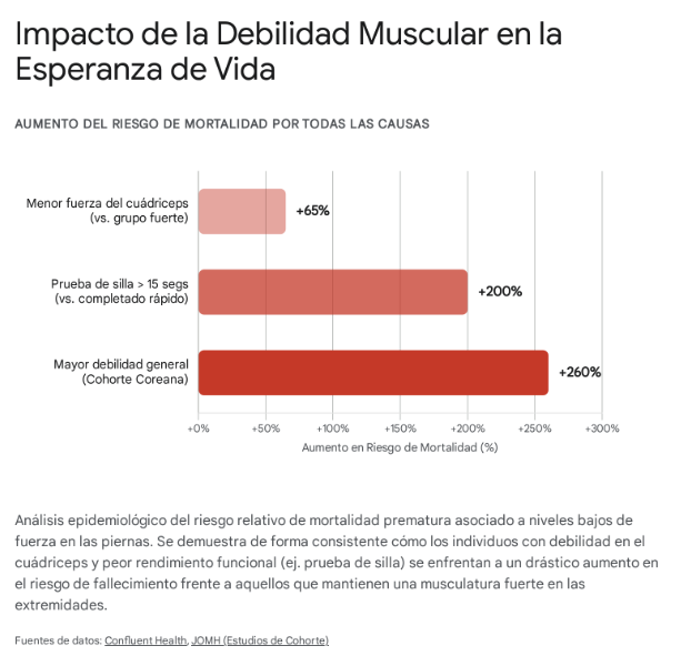
Additionally, the kinematic and functional deficiencies derived directly from this installed structural weakness end up proving that physical autonomy gradually evaporates. The pure, essential capacity of the mature individual to execute everyday autonomous movements, such as repeatedly getting up from a chair, degrades to alarming levels of severe dependence. Under relentless analytical rigor, individuals who took more than 15 seconds to stand up from a chair demonstrated nearly 3 times higher odds of dying prematurely, additionally registering a severe 84% extra probability of needing prolonged hospitalization due to complications from total immobility or accidents compared to vigorous subjects capable of efficiently completing the motor evaluation by achieving the physical challenge.
Added to the viability elements exposed previously against the risk of premature mortality, there is an additional medical constant: strong skeletal muscles in the legs (including calves, quadriceps, and gluteal region) operate by assuming vital metabolic roles. They do not merely act as mechanical support for bipedalism or propulsion against gravity. Due to their immense somatic mass, these peripheral lean tissues function as crucial storage receptacles and unsurpassed biochemical buffers for systemic glucose control in the body.
By operating effectively, these extremities exert their role as indispensable molecular metabolic sinks, controlling glycemic spikes and maintaining balanced endocrine basal metabolism. This profound cellular function halts on a large scale the progressive devastation of chronic hormonal dysfunction, mitigating the incidence of type 2 diabetes mellitus and simultaneously preventing the lethal impact of cardiovascular and coronary conditions at the micro- and macro-arterial levels.
Finally, this muscular vigor in the lower extremities acts as the main biological brake against the pathological involution that ravages old age: sarcopenia. This condition, defined as the terrible, unstoppable loss or gradual wasting of mass, protein volume, and generalized muscle strength, is significantly counteracted by the active maintenance of a solid motor base, defying the irreversible functional effects of the simple chronological passage of aging.
The entire foundational, architectural, biomechanical, and stabilizing base upon which the integrity and firmness of the human body rests non-negotiably resides in the foot and its complex plantar muscle network.
As humans age, the cumulative, prolonged use of restrictive, rigid footwear over decades disproportionately accelerates the wear and cellular degeneration of this natural plantar mechanism. Parametric verification studies in biomechanical laboratories have rigorously contrasted healthy older adults with youth cohorts, demonstrating profound morphological voids and alarming functional deficiencies in the senile population.
There is evidence of an uninterrupted decline encompassing both a decrease in maximum contractile strength and a pathological reduction in the cross-sectional area of the deep stabilizing muscle tissue (atrophy of the abductor hallucis and flexor digitorum brevis). This atrophy severely limits the degrees of joint freedom, diminishing the natural propulsive mechanism and ankle dorsiflexion. Furthermore, countless morphological scans confirm that this atrophy is aggravated by the uncontrolled passive infiltration of intramuscular fatty tissue, which replaces vital contractile cells in the foot’s muscle network.
The intimate correlation demonstrated between this local anatomical atrophy (plantar muscle deficiency) and the general debacle of static and postural balance mechanisms is, today, recognized in geriatric fields as one of the primary epidemiological threats. The loss of strength in these tiny but critical intrinsic foot muscles chains directly to a drastic reduction in the body’s functional limits of spatial stabilization. This weakness joins a dangerous proprioceptive deficiency—a loss of the plantar surface’s neurological sensitivity and agility to read the terrain—that condemns the elderly to a faltering, precarious general motor competence.
These abysmal neuromuscular deficiencies and balance collapses converge into a lethal scourge that threatens all possibility of true longevity and personal autonomy: mechanical falls. Based on rigorous census data, over 30% of adults over 60, and up to a chilling 60% of those over 80, suffer at least one fall per year. These painful, abrupt incidents invariably lead to complex, incapacitating fractures (especially hip and femur breaks), proceeding to inescapable hospitalization, severe immobilization, and triggering marked, irreversible psychological deterioration, social isolation, and total loss of independence.
Restoring the vitality of the plantar base through the proprioceptive reconnection and muscular strengthening facilitated by minimalist footwear stands, therefore, not merely as a biomechanical adjustment, but as a crucial prophylactic intervention to protect skeletal integrity and ensure prolonged functional longevity.
1. The Natural Cushioning and Truss Model (Lapidus Mechanism)
Description: The historical 1943 study published by P.W. Lapidus. In this foundational research, Lapidus demystifies the idea of the foot as a soft spring and proposes for the first time the “truss model,” demonstrating how the bony architecture in conjunction with the plantar fascia mechanically stabilizes the human foot under load.
2. The Cushioning Paradox (Scientific Reports Study)
Title: Highly cushioned shoes increase leg stiffness and amplify impact loading in running
Description: Published in the prestigious journal Scientific Reports (Nature) by Kulmala et al. (2018). This is the exact study cited in your essay that demonstrates that thickly cushioned footwear causes the brain to induce greater leg stiffness, flexing the knees less and paradoxically leading to higher bone impact loading rates than thin-soled minimalist footwear.
3. Minimalist Footwear and “Foot Core” Hypertrophy (5,000 Steps Protocol)
Title: Walking in Minimalist Shoes Is Effective for Strengthening Foot Muscles
Description: A randomized clinical trial led by Ridge et al. (2019) published in Medicine & Science in Sports & Exercise. This study documents how a progressive minimalist shoe walking program (increasing up to 5,000 daily steps) reverses foot atrophy and increases the cross-sectional area of the abductor hallucis (AbH) muscle just as effectively as doing specific clinical rehabilitation exercises.
4. Elevated Heels (Drop), Muscle Shortening, and the Achilles Tendon
Description: A landmark ultrasound investigation by Csapo et al. (2010) in The Journal of Experimental Biology. It analyzes how the constant use of elevated heels alters calf muscle fascicle length and generates a substantial increase in the structural stiffness of the Achilles tendon, permanently altering the range of ankle dorsiflexion.
5. Lower Extremity Strength, Autonomy, and Mortality Risk
Title: Association between muscular strength and mortality in men: prospective cohort study
Description: Large-scale epidemiological research (Ruiz et al., 2008, British Medical Journal) that consolidates the medical evidence presented in your text: the pathological loss of muscle strength and mass in the lower extremities acts as an infallible, direct predictor of increased all-cause mortality, severe sarcopenia, and loss of autonomy in old age.
Research conducted by: Josh Bettencourt
Medical Disclaimer: The mission of Caloritrack (developed and legally operated by KAI STUDIOS, S.A.S.) is to provide you with evidence-based tools and information to take control of your well-being. This in-depth study has been written for purely informational and educational purposes. KAI STUDIOS, S.A.S. is not a healthcare provider. The information presented here should not be interpreted under any circumstance as personalized medical advice, nor is it intended to diagnose, treat, cure, or prevent any disease. Every body is unique; therefore, we strongly urge you to consult with your trusted physician or qualified healthcare professional before implementing new habits, fasting protocols, or metabolic changes based on this study.